
Microscopic examination is routinely utilized in the diagnosis of various dermatological conditions, particularly parasitic and fungal diseases. However, this diagnostic tool is often time-consuming, as it requires patient sampling and specialized equipment. With advancements in technology, imaging methods are rapidly evolving. Recently, there has been an increase in studies focused on smartphone-integrated imaging techniques. For instance, a smartphone-based intraocular lens microscope has been employed in the field of ophthalmology (). Similarly, in dermatology, skin cancers have been assessed using a lens integrated into a smartphone (). We believe that more practical and accessible technologies should be adopted in the field of medicine.
In our study, we utilized a microscopic lens capable of 400x magnification. By integrating this lens into an apparatus designed to function as a phone case and using the smartphone’s camera, we successfully captured images akin to those obtained with a traditional 40x microscope lens. The lens featured not only significant magnification but also an illumination source to enhance the examined area. This innovative approach eliminates the need for patient sample collection and circumvents the expense of using a conventional microscope for microscopic examinations. Additionally, it allows for the capture of photos and videos during the examination of lesions. In our research, we observed the tunnels in scabies patients and examined superficial skin biopsy samples for Demodex evaluation using this smartphone-based microscope. This enabled us to visualize the tunnels, mites, and eggs at a microscopic level (). Through this diagnostic method, we were able to observe the movement of the mites within the tunnels (). When directly examining the superficial skin biopsy sample for Demodex detection with the smartphone, we clearly identified Demodex mites (). We assessed Demodex density by scanning the entire area during preparation ().
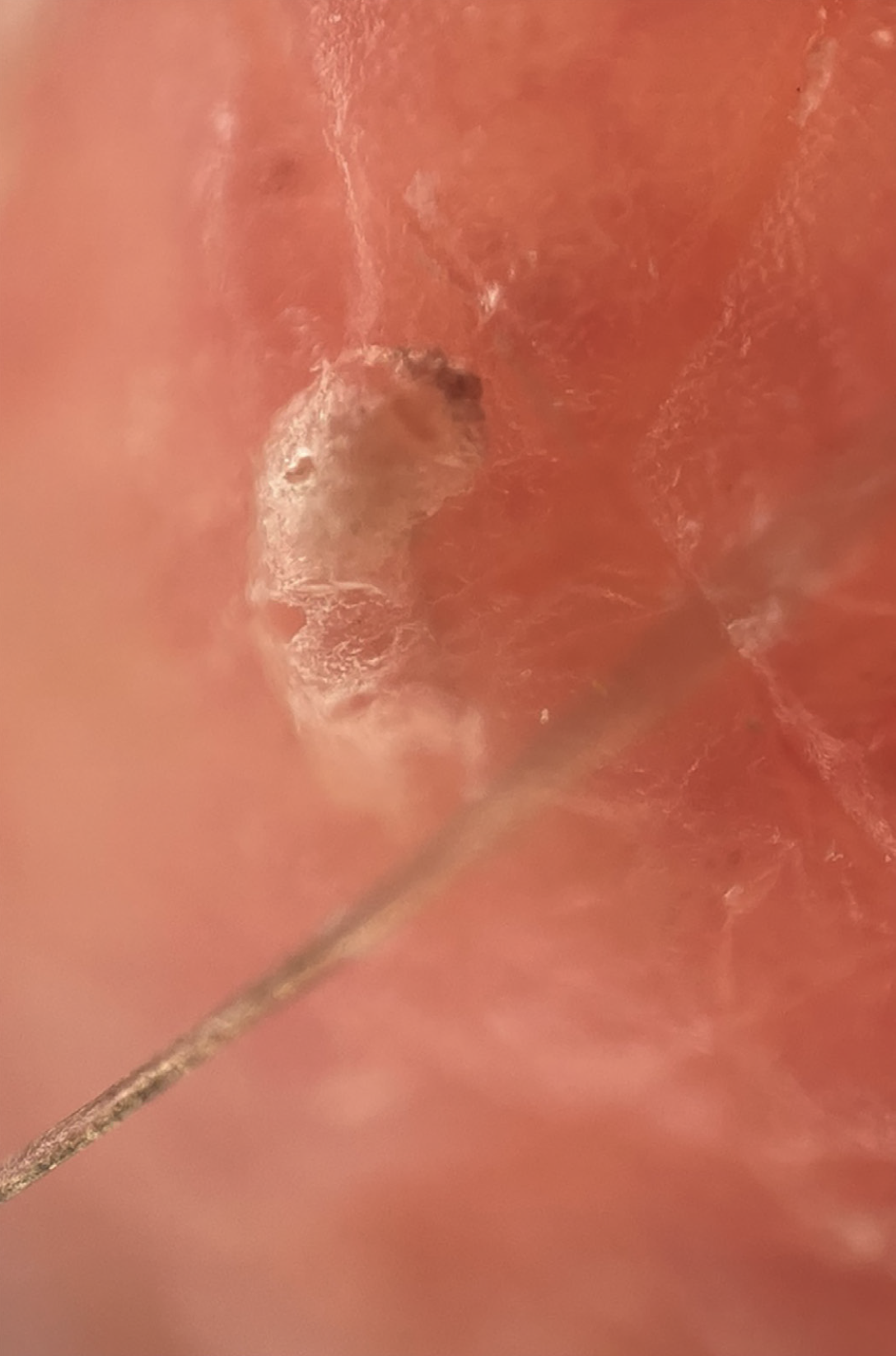
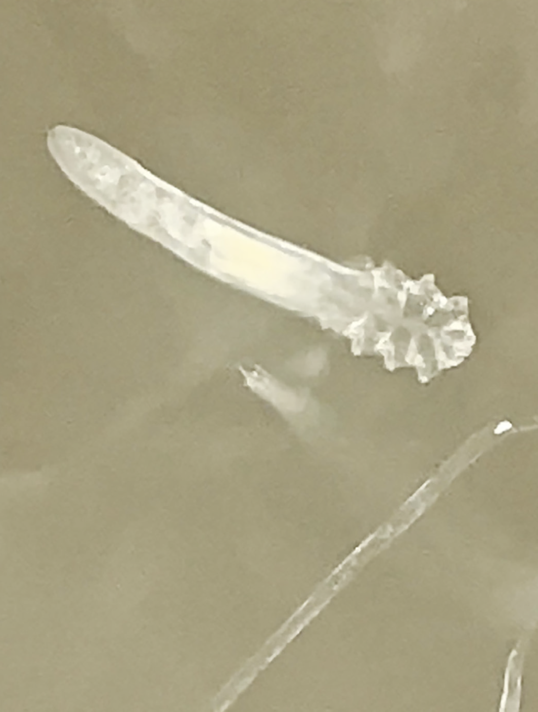
While smartphone manufacturers are continuously enhancing camera features with each new model, the market has yet to offer a lens catering specifically to the medical community. By equipping smartphones with a simple lens capable of 400x magnification, we can effectively meet the significant needs of doctors, biology educators, and microscopy enthusiasts-particularly in dermatology. We believe that our study will greatly facilitate the work of dermatologists and physicians using microscopes in their daily practice while showcasing the potential of advancing technology.
Footnotes
Authorship Contributions
Data Collection or Processing: M.B.A., Literature Search: İ.C., Writing: A.Y.
Conflict of Interest: No conflict of interest was declared by the authors.
Financial Disclosure: The authors declared that this study received no financial support.
References
Chandrakanth P, Chandrakanth KS. Smartphone-based intraocular lens microscope. Indian J Ophthalmol. 2020; 68: 2213-5.
Jahan-Tigh RR, Chinn GM, Rapini RP. A Comparative study between smartphone-based microscopy and conventional light microscopy in 1021 dermatopathology specimens. Arch Pathol Lab Med. 2016; 140: 86-90.